
articles

Dry eye is a common condition that affects millions of individuals worldwide. It occurs when the eyes do not produce enough tears or the tears evaporate too quickly. This can lead to discomfort, irritation, and even vision problems. TearCare offers a breakthrough treatment option for long-lasting relief from dry eye.
What is TearCare?
TearCare is a revolutionary treatment option that offers long-lasting relief for dry eye sufferers. This innovative technology utilizes wearable eyelid warming devices to precisely deliver heat to the meibomian glands, effectively unclogging them and promoting healthier oil production. The treatment is performed in-office by a trained eye care professional and typically takes less than 15 minutes.
TearCare targets the root cause of the problem - meibomian gland dysfunction. By restoring the proper function of these glands, TearCare helps to stabilize the tear film, improve tear production, and alleviate the symptoms of dry eye. This breakthrough solution offers a safe and effective alternative to medications and artificial tear drops, providing long-lasting relief for those suffering from dry eye.
How TearCare Works to Treat Dry Eye
TearCare works by gently applying heat to the eyelids, stimulating the meibomian glands and promoting the release of healthy oils. The wearable eyelid warming devices are designed to maintain a consistent temperature, ensuring optimal results. The heat helps to liquefy any hardened oils or debris clogging the glands, allowing them to function properly again. This treatment can also help reduce inflammation and improve overall eyelid hygiene.

Ocular aesthetics, which focuses on improving the appearance of the eyes and the surrounding area, has gained significant popularity in recent years. Ocular aesthetics aims to rejuvenate the eyes, making them appear more youthful, vibrant, and refreshed. This can be achieved through advanced technology treatments that are designed to address specific concerns and enhance the overall appearance of the eyes.
Common Concerns in Ocular Aesthetics
There are several common concerns that individuals seek to address through ocular aesthetics. These concerns may be a result of natural aging, genetics, or lifestyle factors. One of the most common concerns is the appearance of wrinkles and fine lines around the eyes, commonly known as crow's feet. These lines can make you look older and tired.
Another common concern is the presence of dark circles and puffiness under the eyes. This can give the impression of fatigue and detract from the overall attractiveness of the eyes.
IPL and How it Works
Intense Pulsed Light (IPL) has emerged as a popular and effective treatment option in ocular aesthetics. IPL works by emitting high-intensity light pulses that target specific chromophores in the skin, such as melanin and blood vessels. This targeted approach allows for precise treatment of various concerns around the eyes.
IPL treatments can effectively address pigmentation irregularities, such as age spots and sun damage, which often contribute to an aged appearance. By targeting these concerns, IPL can help restore a more even skin tone and improve the overall aesthetics of the eyes.
IPL treatments are non-invasive and typically require minimal downtime. The number of sessions needed may vary depending on the specific concerns being addressed. By consulting your optometrist, you can determine the best treatment plan to achieve your desired ocular aesthetics.
Radiofrequency (RF) for Ocular Aesthetics
Radiofrequency (RF) treatments have gained significant popularity in ocular aesthetics due to their effectiveness in addressing various concerns around the eyes. RF works by delivering controlled radiofrequency waves into the deeper layers of the skin, stimulating collagen production and tightening the tissues.
One of the primary concerns that RF treatments can address is drooping eyelids. As we age, the skin around the eyes can lose elasticity, causing the eyelids to sag. RF treatments can effectively tighten the skin, resulting in a more youthful and lifted appearance. This can help restore the natural contour and symmetry of the eyes, enhancing their overall aesthetics.
RF treatments can also target under-eye bags, which can make the eyes appear tired and aged. By stimulating collagen production, RF can improve the overall tone and texture of the skin, reducing the appearance of under-eye bags and giving the eyes a more refreshed and vibrant look.
Exploring Low-Level Light Therapy (LLLT)
Low-Level Light Therapy (LLLT) has gained recognition in ocular aesthetics for its ability to promote cellular activity and stimulate collagen production. LLLT utilizes specific wavelengths of light that penetrate the skin, triggering a series of biological responses that can improve the health and appearance of the eyes.
LLLT treatments can effectively address concerns such as fine lines, wrinkles, and dark circles around the eyes. By stimulating collagen production, LLLT can help reduce the appearance of these concerns, resulting in a more youthful and vibrant look.
In addition to its anti-aging benefits, LLLT can also improve the overall health of the eyes. By promoting cellular activity, LLLT can enhance circulation, reduce inflammation, and support the natural healing process. This can be particularly beneficial for individuals with conditions such as dry eyes or eye fatigue.

Clear Lens Extraction (CLE) is a surgical procedure that involves the removal of the natural lens of the eye and replacing it with an artificial lens implant, also known as an intraocular lens (IOL). CLE is primarily performed to correct refractive errors, such as nearsightedness, farsightedness, and astigmatism.
Understanding CLE and How It Works
Clear Lens Extraction is similar to cataract surgery, where the natural lens is removed and replaced with an IOL. However, in CLE, the lens is clear and not clouded as in the case of cataracts. The procedure begins with the administration of local anesthesia to numb the eye and ensure a painless experience for the patient.
Once the eye is numb, a small incision is made on the cornea, which is the clear front surface of the eye. Through this incision, your eye surgeon accesses the lens and carefully removes it. The artificial IOL is then inserted into the empty lens capsule. The IOL is specifically chosen to correct the patient's refractive error, providing them with improved vision.
After the IOL is implanted, the incision is closed with tiny sutures or self-sealing techniques. Following the surgery, patients are usually prescribed antibiotic and anti-inflammatory eye drops to prevent infection and reduce inflammation.
Factors to Consider Before Undergoing Clear Lens Extraction
Before deciding to undergo Clear Lens Extraction, there are several factors that need to be considered. Firstly, it is crucial to have a thorough eye examination to determine if you are a suitable candidate for the procedure. Your optometrist will evaluate your overall eye health and discuss your expectations and goals for vision correction.
Age is another important factor to consider. CLE is typically recommended for individuals over the age of 40 who have developed presbyopia, a condition that affects near vision. It is also important to have stable vision, as any changes in prescription can affect the accuracy of the IOL power calculation.
Additionally, it is essential to understand the potential risks and complications associated with the procedure. While CLE is generally safe, there is a small risk of infection, bleeding, and retinal detachment. Your optometrist will discuss these risks with you and address any concerns you may have.
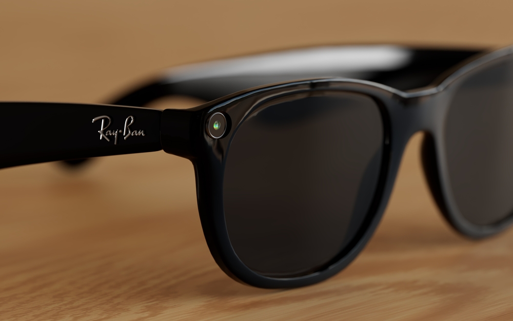
Ray-Ban has long been recognized as a leader in timeless style, but now the brand is taking a bold step into the future. In collaboration with Meta, Ray-Ban has introduced a groundbreaking innovation - Ray-Ban Meta Glasses. These glasses are a fusion of iconic design and next-generation technology that allows you to capture, share, and stay connected like never before.
What Are Ray-Ban Meta Glasses?
Ray-Ban Meta Glasses go beyond vision correction or sun protection - they seamlessly integrate a camera, speakers, and voice-activated AI assistance, allowing users to capture, share, and interact hands-free. With their classic Ray-Ban frames, these glasses look just like traditional eyewear, but they’re packed with cutting-edge features hidden in plain sight.
Key Features and Technology
Built-in Cameras: Capture photos and videos instantly with a simple voice command or tap on the frame. The dual 12MP cameras allow for high-quality media with depth and detail.
Open-Ear Audio: Discreet speakers positioned near your ears provide clear sound without blocking ambient noise, letting you listen to music, take calls, or hear notifications seamlessly.
Meta AI Integration: Access Meta’s AI assistant through voice commands. Ask questions, control your device, or get updates - all without taking your phone out.
Live Streaming and Sharing: Stream directly to social media platforms like Instagram and Facebook in real time, making it easy to share your perspective with your audience.
Touch and Voice Controls: Intuitive touch-sensitive temples and responsive voice commands give you full control over your smart features.
Transitions® Lenses Option: Enjoy lenses that automatically adapt to changing light conditions for optimal comfort both indoors and outdoors.

A tonometer refers to the equipment that is used in tonometry – a test that measures the pressure inside your eyes, also known as intraocular pressure or IOP for short. Tonometry is rarely performed at your average comprehensive eye exam unless you are at high risk of or have been already diagnosed with glaucoma. Fortunately, tonometry can be used to detect changes in eye pressure before they cause any symptoms, enabling prompt action to be taken before your vision is affected.
About Glaucoma
Glaucoma is a common eye condition that occurs when the optic nerve, which connects the eye to the brain, becomes damaged. It’s normally caused by fluid building up in the front part of the eye, which causes the pressure inside the eyes to build. As the pressure increases, the optic nerve becomes increasingly damaged, and this prevents messages from being transmitted between your eyes and brain effectively. As a result, the patient’s vision becomes compromised. Without treatment, the level of vision loss will continue to increase. Unfortunately, any vision that has been lost as a result of glaucoma cannot be restored.
Most of the time, glaucoma develops very slowly which means that many people don’t realize that they are affected until some damage to their vision has already occurred. However, occasionally glaucoma can develop quickly, and symptoms do occur.
Glaucoma symptoms can include:
Red eyes
Intense headaches
Tenderness around the eyes
Eye pain
Seeing rings/halos around lights
Blurred vision
Nausea and vomiting
If you notice any of these symptoms, it’s important that you make an appointment with your eye doctor right away so that you can be assessed. You are likely to have a tonometry test as part of this assessment.

In the realm of eye care, surgical co-management has emerged as a collaborative approach that aims to provide patients with comprehensive and seamless treatment. This concept involves the joint efforts of optometrists and ophthalmologists, each bringing their unique expertise to the table. By working together, these eye care professionals strive to enhance patient outcomes and deliver exceptional care.
The Role of Optometrist and Ophthalmologist in Surgical Co-Management
Surgical co-management is built upon the unique skill sets and areas of expertise of optometrists and ophthalmologists. By understanding their respective roles, you can appreciate the synergy that this collaborative approach fosters.
Optometrists are primary eye care professionals who specialize in the examination, diagnosis, and non-surgical treatment of vision disorders. Their responsibilities in surgical co-management include:
Performing comprehensive eye examinations and evaluations
Monitoring and managing pre-existing eye conditions
Providing pre-operative and post-operative care
Educating patients on surgical procedures and aftercare
Collaborating with ophthalmologists to ensure continuity of care
Ophthalmologists are medical doctors who specialize in the diagnosis, treatment, and surgical management of eye diseases and disorders. Their role in surgical co-management encompasses:
Evaluating patients' candidacy for surgical interventions
Performing complex surgical procedures
Providing specialized medical and surgical care
Collaborating with optometrists to ensure seamless patient care
Monitoring and managing post-operative complications
By combining the expertise of optometrists and ophthalmologists, surgical co-management ensures that patients receive comprehensive and coordinated care throughout their treatment journey.
How Surgical Co-Management Works
Surgical co-management is a well-orchestrated process that involves several key steps. Understanding how it works can help you navigate this collaborative approach with confidence.
Initial Evaluation: The process typically begins with an optometrist conducting a comprehensive eye examination. During this evaluation, the optometrist assesses the patient's visual needs, identifies any potential issues, and determines if a surgical intervention is necessary.
Referral and Consultation: If surgery is recommended, the optometrist refers the patient to an ophthalmologist for further evaluation and consultation. This step ensures that the patient receives specialized medical advice and a thorough assessment of their suitability for the proposed surgical procedure.
Pre-operative Care: The optometrist plays a crucial role in providing pre-operative care, which may include managing any existing eye conditions, ensuring the patient understands the surgical process, and addressing any concerns or questions they may have.
Surgical Procedure: The ophthalmologist performs the necessary surgical intervention, leveraging their specialized training and expertise in surgical techniques.
Post-operative Care: After the surgery, the patient's care transitions back to the optometrist, who closely monitors the recovery process and provides post-operative care and management. This may involve follow-up appointments, monitoring for any complications, and ensuring the patient adheres to the prescribed treatment plan.
Ongoing Collaboration: Throughout the entire process, the optometrist and ophthalmologist maintain open communication and collaborate closely. This ensures that the patient's care is seamless, and any concerns or issues are promptly addressed by the appropriate healthcare professional.

Corneal refractive therapy, also known as CRT, is a simple, painless treatment for refractive eye errors like myopia and has two core benefits. First, it can be used to help patients see clearly during the day without using glasses or contact lenses, giving them the freedom and flexibility that they need to live life to the fullest. Second, CRT has been shown to help slow the progression of myopia, keeping prescriptions under control and potentially reducing the likelihood of patients developing serious eye health problems associated with high myopia in the future.
Here’s everything that you need to know about corneal refractive therapy and what it means for you.
Understanding refractive eye problems
Refractive eye problems like nearsightedness, farsightedness and astigmatism are extremely common, with nearsightedness – also known as myopia – being the most common of all. Patients with myopia can see nearby objects clearly, but those further away become progressively more blurred. Refractive eye errors occur when the shape of the clear dome covering the front part of the eye, called the cornea, impair the light-bending and focusing process in your eyes. This leads to the light ending up in the wrong place inside the eye, and the message that is sent to our brain from our eyes is muddled, causing blurred vision.
What is corneal refractive therapy?
Corneal refractive therapy was initially developed as a treatment to correct and slow the progression of nearsightedness. However, it has also been found to be effective at controlling other refractive errors, including farsightedness, astigmatism and an age-related refractive condition called presbyopia.
CRT is a non-invasive, painless and straightforward method of correcting patient vision so that they don’t need to wear contacts or glasses, and they don’t need laser vision correction surgery to see clearly. CRT uses special contact lenses that are worn overnight and apply light pressure to the cornea in order to reshape it so that light is refracted correctly, and the image sent from the eyes to the brain is clear. The cornea is able to retain this new shape even after the contact lenses are removed the next morning, meaning that you can continue to see clearly for several hours. The more consistently you wear your CRT lenses overnight, the longer your eyes will learn to retain their new shape and eventually, patients can enjoy up to 48 hours of clear vision without using prescription lenses. However, the effects aren’t permanent so if you stop wearing the lenses, your vision will gradually return back to normal over the course of a few days.
Slowing the progression of myopia with corneal refractive therapy
Another key benefit of CRT is that it can actually help to slow the progression of myopia. Most people who are nearsighted find that their eyesight gets progressively worse as they get older. This deterioration may not be rapid, but it can end in patients requiring high prescriptions. Studies have found that patients who have high myopia are more likely to develop serious eye problems in the future, including glaucoma, macular degeneration, cataracts and a detached retina. Regular use of your corneal refractive therapy lenses could help keep your prescription stable and lower your risk of developing these problems.

Dry eye isn’t always just “not enough tears.” Often, the surface of the eye is irritated and inflamed, so tears don’t stay smooth or stable. For some people with more severe symptoms, amniotic membrane therapy can help by acting like a protective bandage that calms the surface and supports healing.
What Are Amniotic Membranes?
Amniotic membranes are derived from the innermost layer of the placenta and are carefully processed for medical use. Amniotic membranes are used to support regeneration of the corneal surface.
They contain growth factors and biologically active components that:
Reduce inflammation
Promote tissue healing
Minimize scarring
Protect the surface of the eye
Because they are immune-privileged and thoroughly screened, they are considered safe and well.
How Do Amniotic Membranes Help Dry Eye?
In moderate to severe dry eye disease, chronic inflammation can damage the corneal surface. This damage can lead to persistent discomfort, fluctuating vision, and delayed healing.
Amniotic membranes work by acting as a biological bandage. Once placed on the eye, they:
Create a protective barrier over the cornea
Reduce surface inflammation
Encourage regeneration of healthy cells
Restore smoother corneal tissue
By addressing the underlying inflammation and surface damage, amniotic membranes do more than simply mask symptoms - they promote healing at the source.

Dry eye is a common condition that occurs when the eyes do not produce enough tears or when the tears evaporate too quickly. There are several factors that can contribute to dry eye, including environmental factors, hormonal changes, and certain medications. Additionally, conditions such as meibomian gland dysfunction and blepharitis can also lead to dry eye.
The Role of Meibomian Gland Dysfunction and Blepharitis in Dry Eye
Meibomian gland dysfunction occurs when the meibomian glands, which are responsible for producing the oily layer of tears, become blocked or do not function properly. This can result in the tears evaporating too quickly and not providing enough lubrication for the eyes. Blepharitis is an inflammation of the eyelids that can cause redness, itching, and a gritty sensation in the eyes. Both meibomian gland dysfunction and blepharitis can contribute to dry eye by disrupting the natural tear film.
Recognizing the Symptoms of Dry Eye
Dry eye can cause a variety of symptoms, which can vary in severity from person to person. Some common symptoms of dry eye include:
Dryness: The most common symptom of dry eye is a feeling of dryness or grittiness in the eyes. This can make it uncomfortable to wear contact lenses or spend long periods of time looking at a screen.
Redness: Dry eye can cause the blood vessels in the eyes to become more prominent, leading to redness and irritation.
Itching: Some people with dry eye may experience itching or a burning sensation in their eyes.
Excessive tearing: Dry eye can also cause excessive tearing. This is the body's response to the irritation caused by the lack of lubrication in the eyes.
If you are experiencing any of these symptoms, it is important to consult with an optometrist for a proper diagnosis and treatment plan.
Neuro-Optometric Rehabilitation
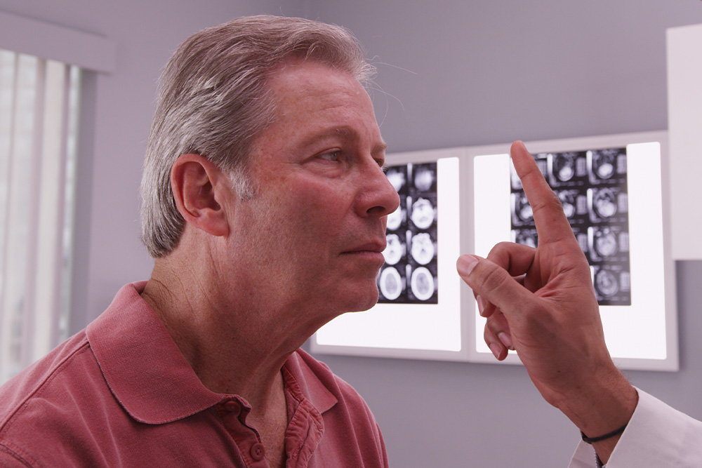
When most people think about vision problems, they picture issues with the eyes themselves. However, many visual difficulties actually begin in the brain. After a concussion, stroke, or other brain injury, the connection between the eyes and brain can be disrupted - causing symptoms that affect balance, reading, focus, and even daily comfort.
Brain Injuries and Vision
Your visual system is complex - about 70% of the brain is involved in vision processing. After a traumatic brain injury (TBI), concussion, or neurological event such as a stroke, the pathways that coordinate eye movement, focus, and perception can become impaired.
This can result in symptoms such as:
Blurred or double vision
Eye strain and headaches
Difficulty reading or focusing
Dizziness or balance problems
Light sensitivity
Problems with depth perception
These visual issues often persist long after other symptoms of a brain injury have improved, making specialized vision care essential for recovery.
What Is Neuro-Optometric Rehabilitation?
Neuro-optometric rehabilitation is a specialized form of vision therapy designed to retrain how the brain and eyes work together. Unlike traditional eye care that focuses on eyesight alone, this therapy targets the neurological processes behind vision. Our doctor evaluates how the eyes and brain communicate and creates a personalized treatment plan to rebuild these visual pathways. The goal is to restore comfort, coordination, and clarity - improving overall visual function and quality of life.


Our Services
Contact Info
Phone: (503) 342-6853
Fax: (503) 342-6185
Hours of Operation
- Monday Closed
- Tuesday 9:00am - 6:00pm
- Wednesday 9:00am - 6:00pm
- Thursday 9:00am - 6:00pm
- Friday 9:00am - 6:00pm
- Saturday 10:00am - 3:00pm
- Sunday Closed
© 2025 Polsinelli Optometry. All rights Reserved. Accessibility Statement - Privacy Policy - Sitemap
Powered by:
